Endovascular technique
Microcatheter navigation provides access to the ophthalmic artery without surgical incisions in the eye.
Geographic atrophy: a new investigational alternative.
Age-related macular degeneration is one of the leading causes of irreversible vision loss in adults over 60. In Buenos Aires, Dr. Mario Joaquín Saravia, President of Fundación Retina, is working on a multidisciplinary ophthalmic artery angioplasty approach.
Geographic atrophy is one of the most severe forms of AMD and can cause progressive loss of central vision.
AMD affects nearly 200 million people worldwide. Current therapies can control or slow some forms of the disease, but they do not restore vision that has already been lost. This is why research into approaches that address other potential mechanisms continues.
One line of research examines the role of vascular supply to the macula. The hypothesis proposes that improving ophthalmic artery blood flow could support ocular and retinal perfusion in carefully selected patients.
Microcatheter navigation provides access to the ophthalmic artery without surgical incisions in the eye.
The procedure is performed under general anesthesia and requires hospitalization and preventive monitoring.
It combines endovascular neurosurgery, retina, and macula expertise for a joint assessment of each case.
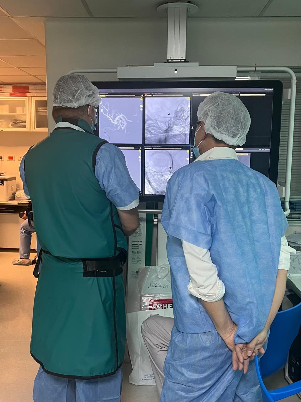 Angiographic visualization during the procedure.
An endovascular procedure designed to restore blood flow in the artery that supplies the eye.
Using a specialized microcatheter, the team accesses the ophthalmic artery, observes the site of obstruction in real time, and uses endovascular devices to open the affected vessel.
A clearly defined process designed around each patient's safety and support.
The team gathers the medical history, ophthalmological studies, and clinical information required for a preliminary evaluation.
In Buenos Aires, the studies provided by the patient are reviewed and the examinations needed to confirm the status of the retina and macula are performed.
MR angiography is used to study the patient's individual vascular anatomy and plan the procedural strategy.
The procedure is performed under general anesthesia. A minimum 24-hour hospital stay is required for observation and preventive monitoring.
After discharge, ophthalmological examinations are repeated, usually one week later, to assess the patient's progress.
The initial protocol was designed to study the feasibility and safety of the procedure.
These figures come from a small pilot series and should be interpreted as preliminary results. They do not establish definitive efficacy, replace medical assessment, or guarantee individual outcomes.
Patients who underwent arterial angioplasty share their experience.
Angioplasty and geographic atrophy patient.
Angioplasty and geographic atrophy patient.
Angioplasty and geographic atrophy patient.
Testimonials reflect individual experiences. They are not evidence of efficacy and do not guarantee that other patients will achieve similar outcomes.
Dr. Saravia leads the ophthalmological assessment, patient selection, and follow-up for each case.
An ophthalmic surgeon specializing in macular and retinal disease. Director of Fundación Retina – Buenos Aires Mácula, with over 28 years of experience in his specialty.
Scientific publications on ophthalmic artery angioplasty for geographic atrophy secondary to AMD.
Get in touch with our team. We will guide you through the documents required and the evaluation process.
For detailed information about the program, patient evaluation, the medical team, scientific updates, and the international patient journey, visit the official treatment website.